.png)
The Anatomy of a Coral Polyp:
A Detailed Exploration
Coral reefs are some of the most diverse and vibrant ecosystems on our planet, and at the heart of these ecosystems are coral polyps. These tiny, soft-bodied organisms form the building blocks of coral reefs, which can stretch for thousands of miles and support an incredible variety of marine life. In this article, we'll dive into the anatomy of a coral polyp, exploring its structure and function in a way that's easy to understand, even if you're just starting to learn about marine biology.
What is a Coral Polyp?
A coral polyp is a small, cylindrical organism that is part of a larger colony of polyps, collectively forming what we commonly refer to as a coral. Each polyp is an individual animal, yet it lives in close connection with other polyps to build the larger coral structure, which is often made of a hard, calcium carbonate skeleton. Coral polyps belong to the phylum Cnidaria, which also includes jellyfish and sea anemones.
Basic Structure of a Coral Polyp

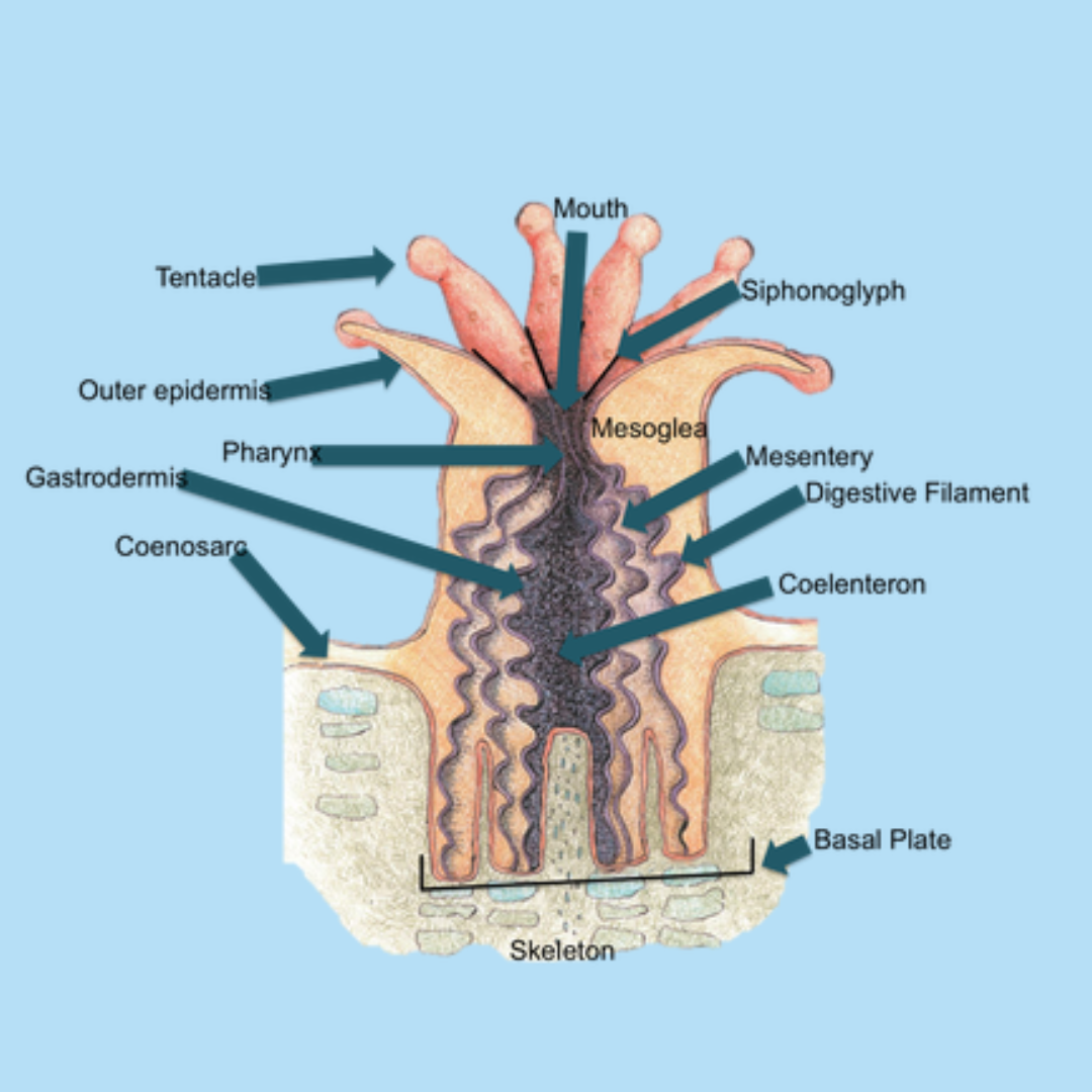
To understand the anatomy of a coral polyp, let's break it down into its main parts:
1. The Mouth
- Description: The mouth is located at the center of the coral polyp’s upper surface, also known as the oral disc. It is usually a small, circular opening that serves as both the entrance for food and the exit for waste.
- Function: The mouth opens to allow the polyp to ingest food, such as tiny plankton or organic particles floating in the water. It also expels waste after digestion.
***Pro-Tip: Imagine the mouth of a coral polyp as a doorway that allows food to enter and waste to leave. Unlike humans, a coral polyp has only one opening for both functions.
2. Tentacles
- Description: Surrounding the mouth are the polyp’s tentacles, which are long, flexible, and often covered in tiny stinging cells called nematocysts. The number of tentacles can vary, but they are usually arranged in a ring around the mouth.
- Function: Tentacles are used to capture food and bring it to the mouth. The nematocysts on the tentacles can sting and paralyze small prey, making it easier for the polyp to consume them. Tentacles can also help in defense against predators and in sensing the environment.
***Pro-Tip: Think of the tentacles as the polyp's "arms," reaching out to grab food and defend itself from threats.
3. Nematocysts (Stinging Cells) .png)
- Description: Nematocysts are specialized cells found on the tentacles and sometimes on the body of the coral polyp. These cells contain a coiled, thread-like structure that can rapidly uncoil and inject venom into prey or potential threats.
- Function: Nematocysts are used for capturing prey by stunning or paralyzing them with venom. They are also an important defense mechanism against predators and competing organisms.
***Pro-Tip: Nematocysts are like tiny harpoons that the polyp uses to catch its prey and defend itself. When triggered, they shoot out to deliver a sting.
4. The Gastrovascular Cavity
-
Description: The gastrovascular cavity, also known as the coelenteron, is the main body cavity of the coral polyp. It is connected to the mouth and functions as both a stomach and a circulatory system.
-
Function: This cavity is where digestion takes place. The polyp secretes digestive enzymes into the cavity to break down food, and the nutrients are absorbed through the cavity walls. The gastrovascular cavity also helps circulate nutrients throughout the polyp’s body.
***Pro-Tip: The gastrovascular cavity works like a simple stomach, where food is digested and nutrients are distributed throughout the polyp.
5. The Epidermis and Gastrodermis
-
Description: The coral polyp has two main layers of cells: the outer layer called the epidermis and the inner layer called the gastrodermis. Between these layers is a jelly-like substance called mesoglea.
-
Epidermis: This outer layer protects the polyp and contains the tentacles and nematocysts.
-
Gastrodermis: This inner layer lines the gastrovascular cavity and is involved in digestion and nutrient absorption.
-
-
Function: The epidermis acts as a protective barrier and plays a role in capturing prey, while the gastrodermis is crucial for digestion and nutrient absorption.
***Pro-Tip: Think of the epidermis as the polyp's "skin" and the gastrodermis as its "stomach lining." Together, they help protect the polyp and digest food.
6. The Calyx (Calcium Carbonate Skeleton)
-
Description: The calyx is the cup-shaped structure made of calcium carbonate that the polyp sits in. This skeleton is produced by the polyp itself and provides structure and protection.
-
Function: The calyx supports the polyp and helps it anchor itself in place. As the coral colony grows, new calyxes are formed, creating the intricate structures of coral reefs.
***Pro Tip: The calyx is like the polyp's "house," providing a sturdy place for it to live and grow. Over time, these calyxes build up to form massive coral reefs.
7. Zooxanthellae (Symbiotic Algae)
-
Description: Zooxanthellae are tiny algae that live inside the tissues of the coral polyp, particularly within the gastrodermis. These algae have a symbiotic relationship with the polyp, meaning both organisms benefit from each other.
-
Function: Zooxanthellae perform photosynthesis, a process that converts sunlight into energy. The polyp uses this energy to grow and build its calcium carbonate skeleton. In return, the zooxanthellae receive protection and access to carbon dioxide and nutrients from the polyp.
***Pro-Tip: Zooxanthellae are like tiny solar panels inside the polyp, providing energy through sunlight. This is why most corals need plenty of light to thrive.
How Does a Coral Polyp Function?
Now that we understand the basic structure of a coral polyp, let’s explore how these parts work together to keep the polyp alive and thriving.
1. Feeding and Digestion
Coral polyps are primarily carnivorous, meaning they feed on small organisms like plankton. The tentacles capture prey and bring it to the mouth, where it is ingested and moved into the gastrovascular cavity. Here, digestive enzymes break down the food, and the nutrients are absorbed by the gastrodermis. Waste products are expelled back through the mouth.
2. Photosynthesis and Energy Production
The zooxanthellae inside the polyp’s tissues perform photosynthesis, using sunlight to produce energy-rich compounds like glucose. This energy is shared with the polyp, allowing it to grow and maintain its calcium carbonate skeleton. This symbiotic relationship is crucial for the health of the coral, which is why corals are often found in shallow, well-lit waters.
3. Protection and Defense
The polyp uses its nematocysts to capture prey and defend against predators. If threatened, the polyp can retract its tentacles and body into the calyx for protection. The calcium carbonate skeleton also serves as a physical barrier against damage and predators.
To learn more about coral defense read our article Here
Why is Understanding Coral Polyp Anatomy Important?
Understanding the anatomy of a coral polyp is essential for anyone interested in marine biology, especially if you’re considering keeping a coral reef aquarium. Knowing how polyps function helps us understand what they need to thrive, such as proper lighting, water quality, and feeding. It also highlights the importance of protecting coral reefs, which are vital to marine life and our planet’s health.
Coral polyps may be small, but they play a huge role in the ocean’s ecosystems. By studying their anatomy, we gain insight into how these amazing organisms live, grow, and build the beautiful coral reefs that so many species depend on.
Conclusion
Coral polyps are incredible organisms that combine complex anatomy with fascinating behavior. Each part of the polyp, from its tentacles to its zooxanthellae, plays a vital role in its survival. By understanding the anatomy of a coral polyp, we can better appreciate the intricate relationships that sustain coral reefs and the importance of protecting these vital ecosystems.
Happy Reefing!
Photo Credit:
- https://courses.lumenlearning.com/
- https://mission31surfaceteam.weebly.com/blog/lets-talk-about-coral
